


Some patients with dementia maintain previously learned board, card-game, painting or musical skills despite progression of their illness (Reference Cummings and ZaritCummings & Zarit, 1987; Beatty et al Reference Beatty, Zavadil and Bailly1988, Reference Beatty, Winn and Adams1994; Reference Edwards-Lee, Miller and BensonEdwards-Lee et al, 1997). We have described patients with frontotemporal dementia (FTD) who developed new artistic skills after the clinical onset of their dementing illness (Reference Miller, Cummings and BooneMiller et al, 1998) and we hypothesised that visual creativity was more common in FTD subtypes with anterior temporal lobe dysfunction. This paper extends those original observations and confirms that visual or musical creativity is present primarily in the subset of patients with predominantly left anterior temporal lobe dysfunction.
METHOD
We reviewed the clinical, neuropsychological and neuroimaging features of every patient (n=69) with a clinical diagnosis of FTD seen at the UCLA Alzheimer's Disease Center. Diagnosis required a compatible clinical syndrome and imaging which showed anterior structural and/or functional deficits as specified in the new research criteria for FTD (Reference Neary, Snowden and GustafsonNeary et al, 1998). We included patients with unilateral left-sided dysfunction presenting with primary progressive aphasia or semantic dementia (Reference Hodges, Patterson and OxburyHodges et al, 1992; Reference Snowden, Neary and MannSnowden et al, 1996). Age and psychiatric status were not inclusion or exclusion factors and dementia severity varied. All received behavioural, psychological and imaging as previously described (Reference Miller, Cummings and BooneMiller et al, 1998). Care-givers were queried about preservation of skills such as language, navigation, painting, music, card and board games.
Neuropsychological tests were designed to assess executive skills, language, visuoperception, visuo-construction and memory (Reference Pachana, Boone and MillerPachana et al, 1996). Magnetic resonance imaging (MRI) was used to exclude focal brain lesions. Single photon emission computed tomography (SPECT) was performed with 133Xe, which gave an absolute measure of regional cerebral blood flow, and 99Tc-labelled hexamethyl-propyleneamineoxime (HMAO), which provided high resolution qualitative images.
Two types of statistical comparisons were performed. First, we compared the 12 patients with creative skills (with ability) with 12 Mini-Mental State Examination (MMSE)-matched (Reference Folstein, Folstein and McHughFolstein et al, 1975) subjects with FTD (without ability) on tests we find best for distinguishing asymmetrical left-sided from right-sided FTD (Reference Boone, Miller and LeeBoone et al, 1999). These include the Boston Naming Test (Reference Kaplan, Goodglass and WeintraubKaplan et al, 1983) (lower with left-sided lesions), verbal IQ minus performance IQ (Reference Adams, Smigielski and JenkinsAdams et al, 1984) (higher with right-sided lesions), and ‘D-word’ minus design fluency (Reference LezakLezak, 1976) (higher with right-sided lesions). For ‘D-word’, patients were allowed one minute to name words that begin with the letter D. For design fluency, patients used one minute to make novel designs by connecting four dots with straight lines within a box with five dots. The Neuropsychiatric Inventory (NPI) allow measurement of psychiatric symptoms (Reference Cummings, Mega and GrayCummings et al, 1994).
Also, we compared SPECT scans from the subjects with ability with those from the entire FTD cohort (in total, 57 SPECT scans were available). Scans were classified as bilateral, left-, or right-sided by two clinicians blind to patient identity. Patients were noted who had anterior temporal lobe hypoperfusion but sparing of dorsolateral frontal cortex, the temporal lobe variant of FTD (Reference Edwards-Lee, Miller and BensonEdwards-Lee et al, 1997). Perfusion patterns in the subjects with ability v. the entire FTD population were compared.
RESULTS
Preserved creativity in patients with visual skills learned earlier in life
Patient 1: A 74-year-old left-handed female was referred for assessment of progressive aphasia. Most of her life she showed a mixture of scientific proficiency and shyness. A brilliant inventor, she had exceptional visuo-spatial abilities, successfully designing a chemical detector which she perfected throughout life. Yet she never enjoyed reading, and was shy. Language difficulties began at age 68 years, and by 71 she began to lose the meaning of words. She named objects by the superordinate category, calling most machines “instruments” and food items “ materials”. She stopped reading, but continued to perfect her inventions, receiving new patents for innovations during her 74th year. She became withdrawn, but disinhibition emerged. She became hypochondriacal and an aversion to meat persisted. On examination she was pleasant and cooperative but passive. Her MMSE score was 21. The only points lost on the MMSE were related to language. The Rey-Osterrieth Complex Figure copy was normal, and visual memory for the item near normal. She generated six D-words, and 15 novel designs. Comprehension was good for two-step commands. She was profoundly anomic obtaining 1/60 words on the modified Boston Naming task. She read well except for words that did not follow regular phonetic rules (gnat or yacht). A SPECT revealed bitemporal, left greater than right, hypoperfusion with frontal sparing.
Patient 2: A 70-year-old right-handed woman was referred for assessment of progressive aphasia. She spoke eight languages, and played bridge at a professional level. Symptoms began around age 64 when she noticed difficulty with memory. On IQ testing at age 68, arithmetic was in the 99th percentile, comprehension the 50th percentile and general information the 16th percentile. Verbal learning delayed recall was in the 1st percentile. The Boston Naming test was at the 59th percentile. Visual reproduction abilities and the Wisconsin Card Sort Test were normal. Focal left temporal lobe atrophy was apparent on MRI. Word-finding trouble increased, but her visual and social skills remained intact and she continued playing bridge and chess. She became obsessed with playing computer chess and solitaire and considered suicide. Her MMSE score was 25. She missed three recall items, could not remember the name of the city and called a watch a key. Her digit span was eight forward. She could follow two, but not three-step commands and could not name bed or flower. Visual memory recall was impaired, but recognition was good. She generated two D-words and nine novel designs. Her SPECT showed bitemporal hypoperfusion, left greater than right, with frontal sparing.
Patient 3: A 76-year-old right-handed male was referred for assessment of a progressive five-year language and memory disorder. Previously an aerospace engineer, he designed aeroplanes and owned patents for items he had invented. Memory worsened and he developed slurred speech and decreased speech volume. Because of apathy he stopped pursuing hobbies. Navigation skills remained normal, but he was highly productive related to visual talents and successfully designed and built a lighting system and a garage. On examination he was cooperative but placid. His MMSE score was 22. He missed points for recall, orientation and repetition. Verbal IQ testing showed comprehension in the 2nd percentile and arithmetic in the 84th percentile. His performance IQ was 108 with picture completion in the 95th percentile, picture arrangement in the 99th percentile and block design the 84th percentile. He named 16/60 words on the Boston Naming Test. Controlled oral word association was in the 2nd percentile, verbal memory <1st percentile. He did well on most frontal systems tasks. A SPECT showed symmetrical bitemporal hypoperfusion with frontal sparing.
Musical ability which developed in the setting of dementia
Patient 4: A 49-year-old, right-handed man developed progressive aphasia. Previously gifted at foreign languages, he obtained an MS in linguistics. Subsequently, he worked as a mechanic. At age 42 he became withdrawn. His diet changed and meals consisted of chips and sweets. He constantly whistled, mastering classical and popular pieces. He composed musical songs about his bird. At work he became known as ‘the whistler’. Previously shy, he became inappropriately exuberant. At age 47 naming and memory problems emerged and he covered his house with reminder notes. He was forced to retire owing to odd behaviour and moved in with his brother. He became childlike and euphoric, constantly whistling and singing songs about his bird. A compulsive tendency to use the bathroom emerged. His MMSE score was 17. On the Boston Naming Test he named 4/60 items. Memory testing was impaired but he easily learned the floor structure of the medical office building. He generated two D-words but nine novel designs. Complex designs were copied perfectly and he performed well on the Modified Trails. SPECT showed left greater than right temporal hypoperfusion with frontal sparing.
Patient 5: A 78-year-old right-handed man began to mix up the names of his children, dogs and horses. He received little formal education but was a gifted linguist, learning Chinese, Italian and several Russian languages with an accent that led him to be mistaken for a native speaker. He enjoyed playing word puzzles because they allowed him to “understand the psychology of the designer”. Despite receiving limited musical training, at 68 years began to compose classical music. His mind was ‘taken over’ during composition, and he ‘sensed’ different tonal intervals that he put to music. Some pieces were publicly performed and composing continued even after his language and puzzle skills decreased. On examination he was stooped, quiet and remote. His MMSE score was 25. Verbal output was decreased with word-finding circumlocutions. He could not remember the name of any compositions that he had performed and remembered only one of three words after three minutes, but described recent events with accuracy. A SPECT showed moderate left temporal and mild left frontal hypoperfusion.
Previously developed musical and visual ability which persisted in the setting of dementia
Patient 6: A 71-year-old right-handed male was seen five years after the onset of aphasia. At 66 he became quieter and by 69 was exhibiting word-finding difficulty. At 70 reading and writing ceased and comprehension diminished. A talented musician, he could compose songs that captured the personality of acquaintances. He continued public concerts until age 71; only in the year prior to evaluation did this skill deteriorate. An excellent chess and crossword puzzle player, during the two years prior to evaluation his interest in these games increased. At evaluation he still paid bills, and successfully navigated. On examination his MMSE score was 15. Digit span was six. Speech was fluent with many verbal perseverations. He named 4/15 objects and followed two-step commands. He failed formal verbal and non-verbal testing, but remembered the participants in a professional basketball game from two days earlier. No D-words and one design was generated. He could add 389 and 676. Positron emission tomography showed selective left anterior temporal hypometabolism.
(Patients 7-11 developed new skills as visual artists and patient 12 showed preservation of visual and musical skills. All were previously described (Reference Edwards-Lee, Miller and BensonEdwards-Lee et al, 1997; Reference Miller, Cummings and BooneMiller et al, 1998). Their neuropsychological and SPECT data are included here for analysis.)
Statistical analyses
Statistical testing was performed with Fisher's exact test and Student's t-test. Table 1 shows the age at onset of FTD the age when the abilities emerged, the type of ability and the location for the anatomical dysfunction suggested by SPECT (or positron emission tomography in Patient 6, and pathology in Patient 8) is described. Seven developed new skills (five visual and two musical) in the setting of dementia, while five maintained visual and/or musical abilities (two visual, one musical, two both visual and musical) despite progression of dementia. Nine of the 12 patients with ability showed left-sided predominant hypoperfusion. Only 12 of the 45 patients without ability were left-sided. This difference was highly significant using Fisher's exact test (χ2=7.55; P <0.006). Eight of 12 subjects with ability had a temporal lobe variant pattern. In the remaining subjects with FTD without ability (45 in total), nine showed the temporal lobe variant. This difference was highly significant using a two-tailed Fisher's exact test (P<0.0035).
Table 1 Clinical and imaging features of creative patients with frontotemporal dementia
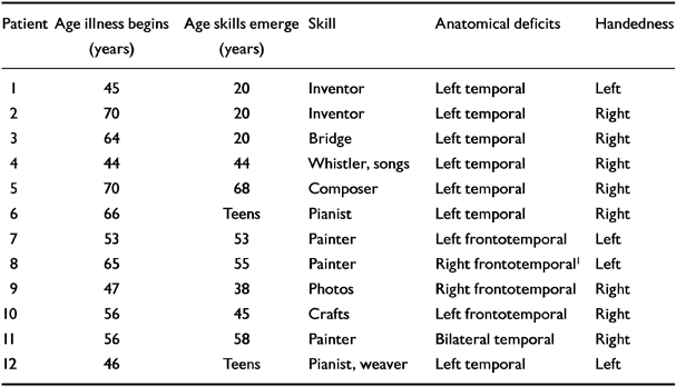
| Patient | Age illness begins (years) | Age skills emerge (years) | Skill | Anatomical deficits | Handedness |
|---|---|---|---|---|---|
| 1 | 45 | 20 | Inventor | Left temporal | Left |
| 2 | 70 | 20 | Inventor | Left temporal | Right |
| 3 | 64 | 20 | Bridge | Left temporal | Right |
| 4 | 44 | 44 | Whistler, songs | Left temporal | Right |
| 5 | 70 | 68 | Composer | Left temporal | Right |
| 6 | 66 | Teens | Pianist | Left temporal | Right |
| 7 | 53 | 53 | Painter | Left frontotemporal | Left |
| 8 | 65 | 55 | Painter | Right frontotemporal1 | Left |
| 9 | 47 | 38 | Photos | Right frontotemporal | Right |
| 10 | 56 | 45 | Crafts | Left frontotemporal | Right |
| 11 | 56 | 58 | Painter | Bilateral temporal | Right |
| 12 | 46 | Teens | Pianist, weaver | Left temporal | Left |
Table 2 summarises the pertinent neuropsychological findings. Average score on verbal IQ minus performance IQ was ‒15.0 (s.d.=9.9) in the ability group and +4.4 (s.d.=9.2) in the group without ability (t=4.4; P=0.0004). Average D-words minus visual designs was ‒5.8 (s.d.=5.2) in the group with ability v. +2.7 (s.d.=2.7) in the group without ability (t=4.6; P=0.0002). The Boston Naming mean scores were 20.3 (s.d.=21.6) in those with ability and 46.5 (s.d.=16.0) in those without ability (Student's t=3.1; P=0.006). These results suggest more left-sided dysfunction in the group with ability. Table 3 summarises neuropsychiatric features. Both groups scored highly on the disinhibition, apathy and aberrant motor behaviour scales. The only statistically significant item was the depression scale; higher in the group with ability (P=0.023).
Table 2 Neuropsychological features of creative subjects with frontotemporal dementia

| Patients | Years since creativity | Mode of creativity | MMSE | Modified Rey-0 copy | D/design fluency | VIQ-PIQ | Boston Naming |
|---|---|---|---|---|---|---|---|
| 1 | Current | Inventing | 21 | Normal | 6/15 | -30 | 1/60 |
| 2 | Current | Inventing | 22 | Normal | 0/12 | -24 | 6/15 |
| 3 | Current | Bridge | 25 | Normal | 2/9 | -17 | 4/60 |
| 4 | Current | Limericks | 17 | Normal | 2/9 | -12 | 4/60 |
| 5 | Current | Piano | 15 | Normal | 0/1 | - | 6/15 |
| 7 | One | Painting | 16 | Normal | 2/7 | -20 | 0/60 |
| 9 | Five | Photos | 26 | Normal | 6/7 | +4 | 50/60 |
| 10 | Five | Crafts | 9 | Normal | 0/1 | -8 | 1/15 |
| 11 | Current | Painting | 15 | Normal | 2/17 | -10 | 32/60 |
| 12 | Current | Piano, weaving | 1 | Normal | 0/0 | -18 | 0/15 |
Table 3 Neuropsychiatric features of creative subjects with frontotemporal dementia

| Patient | Mode | Compulsion | Depression | Family HX1 | Premorbid strengths |
|---|---|---|---|---|---|
| 1 | Inventing | Picks teeth, spits | No | Yes | Brilliant scientist |
| 2 | Inventing | None | No | Yes | Scientist, inventor |
| 3 | Plays bridge | Solitaire | Yes | Yes | Multilingual, excellent game player |
| 4 | Whistler, songs | Toileting, eating | No | Yes | Multilingual |
| 5 | Music composer | Unknown | No | Yes | Multilingual |
| 6 | Pianist, chess | Crosswords, chess | Yes | Yes | Pianist, bridge player |
| 7 | Painting | Searches for coins | No | Possible | None known |
| 8 | Painting | None | Yes | Yes | Intelligent |
| 9 | Photographs | Collects wax | Yes | Yes | Advertising designer |
| 10 | Handicrafts | Folds plates | Yes | Yes | Artistic |
| 11 | Painting | Searches for coins, dietary fads | Yes | Possible | Memory for stocks |
| 12 | Plays piano, weaves | Word games, copies Bible, dietary | Yes | No | Pianist and weaver |
DISCUSSION
Clinical overview
We describe the clinical features of 12 patients with FTD with new or preserved musical or visual ability. These remarkable patients represent 17% of our population with FTD, suggesting that musical or visual creativity is not rare with this dementia syndrome. Analysis of the clinical, neuropsychological, neuropsychiatric and neuroimaging features of these patients suggests an anatomical correlate for musical or visual talent in FTD.
Despite the diversity of visual and musical talents exhibited in this population, they shared many features. Talents were musical or visual and never manifested in the verbal sphere. Work lacked a symbolic or abstract component, and painters copied or remembered realistic landscapes, animals or people or perfected visual designs. A bridge champion and a chess master continued playing these games by manipulating visual images of cards or chess pieces learned earlier in life. For musicians, notes came automatically as sounds without conscious verbal manipulation. These processes have in common the recall of previously learned information or images. Subsequently this information is manipulated without the mediation of language.
Despite progressive cognitive and social impairment, patients showed increasing interest in the fine detail of objects, shapes, sounds, and visual or musical patterns. All neglected social and occupational responsibilities to pursue creative activities and their interests became progressively more restricted. Painting, photography, inventing, bridge, chess, composing, or music became obsessive preoccupation, and this obsessive interest with frequent repetition of the creative activity was an important factor in the development of a quality product.
Anatomical/physiological/neurochemical considerations
Frontotemporal dementia causes selective anterior frontal and/or temporal degeneration, whereas in Alzheimer's disease, early pathology involves entorhinal and posterior parietal and temporal areas (Reference BrunBrun, 1993). In many patients with Alzheimer's disease artistic and musical skills dissipate rapidly (Reference Cummings and ZaritCummings & Zarit, 1987) associated with diminished function in posterior parietal and temporal regions, brain areas that contribute to visuoconstructive, linguistic and musical skills (Reference BrunBrun, 1993). However, in some patients with Alzheimer's disease previously learned musical skills remain intact despite progressive loss of other cognitive functions (Reference Beatty, Winn and AdamsBeatty et al, 1994).
In contrast to the decline in visuo-spatial skills characteristic of Alzheimer's disease, patients with FTD often retain visuo-constructive abilities as pathology is lacking in posterior parietal regions (Reference BrunBrun, 1993). Sparing of these areas allowed our subjects to paint, build, weave and play or compose music, despite advancing dementia. Yet, even though visuo-constructive skills are preserved in many patients with FTD, diminished, not enhanced, creativity is the rule with FTD (Reference Snowden, Neary and MannSnowden et al, 1996), suggesting that individuals who maintain or develop new creative skills have a distinctive form of this disorder.
Eight had the temporal lobe variant of FTD (Reference Edwards-Lee, Miller and BensonEdwards-Lee et al, 1997), which represents almost 50% (8/17) of our patients with this anatomical subtype. Furthermore, another three had a temporally predominant imaging or perfusion deficit even though the frontal lobes were involved by the time of our evaluation. Also, all seven subjects with ability studied with SPECT when talents were present showed a pure temporal lobe perfusion deficit. In contrast, in patients with FTD without ability, and in the patients with ability whose productivity had ceased, widespread frontal and temporal hypoperfusion was seen with SPECT. Involvement of the temporal but sparing of frontal regions was a unifying feature of the patients with ability.
Organisation and motivation are mediated via dorsolateral and medial frontal areas. Goldman-Rakic (Reference Goldman-Rakic1996) observed that: “ Visuospatial processes engaged in humans by activities such as chess playing, following maps, recalling one's location with respect to landmarks, or painting and drawing from memory… rely on the dorsolateral prefrontal convexity…”
Sparing of working memory (Reference Waltz, Knowlton and HolyoakWaltz et al, 1999) and episodic memory (Reference Hodges, Patterson and OxburyHodges et al, 1992) characterise the temporal variant of FTD and probably aided in the musical and visual creativity. Organising a musical piece or painting requires working memory. Similarly, the painters' capacity to draw scenes remembered from the past, and the game players' and musicians' ability to recall previously learned visual and musical patterns suggested that components of episodic memory were spared. In contrast, semantic memory, which requires the integrity of the left anterior temporal lobe (Reference Hodges, Patterson and OxburyHodges et al, 1992; Reference McCarthy and WarringtonMcCarthy & Warrington, 1994; Reference Snowden, Neary and MannSnowden et al, 1996), was impaired.
Most of the patients in the group with ability showed asymmetrical left hemisphere degeneration, which is congruent with the studies suggesting that left hemisphere injury does not invariably decimate musical skills. The composers Shebalin and Langlais suffered strokes in the left temporal-parietal regions which, despite inducing aphasia, did not negatively influence note reading or musical composing abilities (Reference SergentSergent, 1993), and Ravel, who suffered from a focal degenerative disorder of the left hemisphere, continued composing while aphasic and agraphic (Reference SergentSergent, 1993). Polk & Kertesz (Reference Polk and Kertesz1993) described a patient with progressive aphasia in whom “organized, although reiterative music production” continued despite aphasia progression. Their patients were reminiscent of Patients 4-6 and 12 in whom music production was possible despite progressive aphasia. In contrast, McFarland & Fortin (Reference McFarland and Fortin1982) described an accomplished organist who lost his ability to play familiar melodies following infarction of the right superior temporal and supramarginal gyri. The right hemisphere is dominant for spontaneous production of music (Reference Gordon and BogenGordon & Bogen, 1974) and was spared (if not enhanced) in our subjects.
Right hemisphere injury devastates the ability to copy, or paint, even in experienced artists (Reference Schnider, Regard and BensonSchnider et al, 1993) while left hemisphere injury does not routinely injure painting skills (Reference AlajouanineAlajouanine, 1948). Kaczmarek (Reference Kaczmarek1991) described a painter who suffered a left hemisphere stroke with aphasia who lost the ability “to create the highly symbolic pictures he used to paint before the stroke”. Yet, he continued to draw realistically. Kaczmarek noted that there was “ impairment of symbolic processing with fairly well preserved technical skills…” Realistic reproduction of music, or paintings without symbolism was a feature of the creativity in our cohort.
Paradoxical functional facilitation
To explain the unexpected occurrence of behavioural improvement following brain injury, Kapur (Reference Kapur1996) used the term “paradoxical functional facilitation”. He noted that “ in normal subjects, inhibitory and excitatory mechanisms interact in a complex harmony…. The role of inhibitory processes may be critical in mediating specific restorative paradoxical functional facilitation effects”. Paradoxical functional facilitation may have contributed to the unexpected emergence of talent in our patients.
The temporal gyri process visual and auditory information (Reference Baylis, Rolls and LeonardBaylis et al, 1987) and specific cells in the inferior temporal gyrus respond to distinctive visual stimuli including shape and movement. There are many connections to and from polar temporal cortex, and posterior temporal cortex. The role of anterior temporal cortex in processing of visual and musical information is unknown, but enhanced artistic skill in patients with anterior temporal and orbital frontal degeneration suggests that these areas inhibit dorsolateral frontal and posterior temporal and parietal regions involved with these processes.
Paradoxical increases in brain perfusion occur in patients with focal cerebral lesions, (Reference Weiller, Chollet and FristonWeiller et al, 1992), whereas visual stimulation reduces blood flow in prefrontal regions (Reference Kawashima, O'Sullivan and RolandKawashima et al, 1995). Patient 11 described painfully intense visual images, suggesting abnormally intense experiencing of visual images. Patients 7 and 9 found coins with uncanny ability. At age 68, patient 10 suddenly had musical patterns which triggered musical composition. We hypothesise that selective degeneration of left anterior temporal cortex led to decreased inhibition of the more right-sided and posteriorly located visual and musical systems, and dorsolateral frontal regions involved with working memory, thereby enhancing artistic interest and productivity.
Often, talent in one area is accompanied by dysfunction in other spheres and unusual brain organisation may have been important in this subgroup. Four were left-handed and, as a group, they were exceptionally gifted in linguistic, musical or visual tasks prior to the onset of their dementia. The two inventors showed life-long visual talent, yet both were socially withdrawn. Brain asymmetries (possibly congenital) may have predisposed to talents that developed or persisted. Some exhibited lifelong eccentricity suggesting that the illness began early in life and progressed with remarkable slowness. Did loss of function in the left anterior temporal lobe lead to brain remodelling and consequent enhancement in function in the right hemisphere or posterior parts of brain necessary for production of music and art?
Neuropsychiatric features and influences
On the NPI, the group with ability had greater disinhibition than the group without ability, although this difference did not reach statistical significance. Also, disabling compulsions were common in our patients. Two artists searched for coins, a photographer crafted candle wax into miniature animals, a craftsman folded paper plates, a bridge player and pianist repetitively played solitaire and chess for hours at a time, and a weaver and pianist compulsively copied from the Bible and played ‘search and circle’ games. Their artistic achievements were, in part, attributable to their compulsive repetition of musical or visual tasks.
Psychiatric illness is common among artists and Post (Reference Post1994) found that depression was more common than expected among writers, whereas “severe personality deviations” were more frequent among visual artists. Depression was more common in our patients with ability although the relationship between the depression, disinhibition and compulsions and the emergence of creativity is unknown.
Clinical Implications and Limitations
CLINICAL IMPLICATIONS
-
▪ Selective degeneration of the left anterior temporal lobe is often accompanied by loss of semantic knowledge, but preservation or enhancement of visual and musical abilities.
-
▪ Visual and musical abilities should be encouraged in the setting of left anterior temporal injury or dysfunction.
-
▪ Dementia is not invariably associated with relentless loss of all intellectual abilities.
LIMITATIONS
-
▪ The data in this study were analysed retrospectively.
-
▪ Some of the individuals in this study were highly intelligent prior to the onset of their symptoms, suggesting that the emergence of talent may occur only in select patients.
-
▪ The physiological basis underlying the ability to develop new artistic functions in the setting of dementia remains unknown.


eLetters
No eLetters have been published for this article.