


Eating disorders, which have been associated with concerns about body shape and size (American Psychiatric Association, 1994), are about 10 times more common in women than in men (Reference Weissman and OlfsonWeissman & Olfson, 1995). A possible reason for this difference in susceptibility might be a gender difference in the neural processing of unpleasant information about body image. We previously reported that women showed amygdalar activation while processing unpleasant words concerning body image and perceived these words to be emotionally negative (Reference Shirao, Okamoto and OkadaShirao et al, 2003a ). The medial prefrontal cortex has connections to the amygdala, constituting an interaction zone between emotional and cognitive processing (Reference Drevets and RaichleDrevets & Raichle, 1998). In this study we compared the brain activation between men and women while processing these words. We predicted that the amygdala would be less activated and the medial prefrontal cortex more activated in men than in women during the emotional decision task.
METHOD
Study sample
An age-matched sample of 13 men (mean age 25.3 years, s.d.=2.8, range 21–30) and 13 women (mean age 25.2 years, s.d.=3.2, range 21–30) participated in this study (P=0.949 by two-tailed, two-sample Student's t test). Participants were recruited by community announcement and paid incentives equivalent to their transportation expenses. All of them were right-handed and were native Japanese speakers. Handedness was determined using the Edinburgh Handedness Inventory (Reference OldfieldOldfield, 1971). According to self-report, participants had no history of psychiatric, neurological or other major medical illness, and had never been treated with a psychotropic medication. There was no significant difference in the average years of education between men and women: men 15.2 (s.d.=1.6) v. women 14.9 (s.d.=2.5); P=0.645 by two-tailed, two-sample Student's t-test. The average body mass index of the men was 22.4 kg/m2 (s.d.=3.2, range 18.0–31.3) and that of the women was 21.5 kg/m2 (s.d.=3.7, range 18.8–28.4); P=0.543 by two-tailed two-sample Student's t-test. The average of the total Eating Disorder Inventory – 2 (EDI–2; Reference GarnerGarner, 1991) scores of men was 45.5 (s.d.=28.4, range 9–103) and that of women was 37.9 (s.d.=23.5, range 7–85); P=0.330 by two-tailed Wilcoxon single-rank test. The average score for the item ‘body dissatisfaction’ for the men was 7.43 (s.d.=5.45, range 2–19) and for the women was 11.31 (s.d.=7.00, range 0–22); P=0.330 by two-tailed Wilcoxon single-rank test. The study was conducted using a protocol approved by the ethics committee of Hiroshima University School of Medicine. All individuals provided written informed consent for participation in the study.
Emotional decision task
We used the emotional decision task developed by Tabert et al (Reference Tabert, Borod and Tang2001), with some modifications. The words used in the task were selected from the database of Toglia & Battig (Reference Toglia and Battig1978), which includes 2854 words that have been rated on several items such as familiarity and pleasantness, on a scale of 1 (very unfamiliar; very unpleasant) to 7 (very familiar; very pleasant), with 4 as the mid-point. For our study, 30 neutral words were selected from the database and translated into Japanese. We also selected 30 highly unpleasant words concerning body image, chosen from Japanese-language dictionaries and thesauri. The two groups of words did not significantly differ with regard to word length (mean length in Japanese letters: body image words 3.2, neutral words 3.1; P=0.575 by two-tailed, two-sample Student's t-test). Our previous validation study comparing women who had eating disorders with a control group of healthy women showed that there was no significant difference in familiarity between the two categories of words (eating disorder group mean familiarity score: body image words 4.2; neutral words 4.1, P=0.727; control group mean familiarity score: body image words 3.9, neutral words 4.1, P=0.218, by two-tailed Wilcoxon single-rank test) and there was no significant difference in the familiarity ratings of words concerning body image between women with eating disorders and the control group (P=0.365 by two-tailed Wilcoxon single-rank test), whereas there were significant differences in pleasantness between the two categories of words (mean pleasantness score in the eating disorder group: body image words 2.4, neutral words 3.9, P=0.0002; mean pleasantness score in the control group: body image words 3.0, neutral words 4.0, P=0.0001, by two-tailed Wilcoxon single-rank test) and there were significant differences in the ratings of pleasantness between the eating disorders group and the control group (P=0.030 by two-tailed Wilcoxon single-rank test) (Reference Shirao, Okamoto and OkamotoShirao et al, 2003b ). Both lists of words contained nouns, verbs, adjectives and adverbs.
The selected words were used to generate sets of unpleasant words concerning body image and sets of neutral words. Each word set comprised a unique combination of three words. The word sets were presented in six alternating blocks of two conditions (the task condition and the control condition) in three cycles (Fig. 1). During the task condition unpleasant word sets were presented, and during the control condition neutral word sets were presented. Each block began with a 3 s cue identifying the condition by displaying the word ‘task’ or ‘control’. Five word sets were presented in each block. Each word set was shown for 4 s with a 1.4 s interstimulus interval (Fig. 1). The blood oxygen level-dependent (BOLD) response was recorded during three blocks of unpleasant words and three blocks of neutral words. During each interstimulus interval, a fixation cross placed centrally on the screen replaced the word set. Baseline functional magnetic resonance images were obtained during a 9 s period prior to the first block of trials, during which the individual viewed a centrally placed fixation cross. During each trial, the word set was projected to the centre of the person's field of view by a Super Video Graphics adapter computer-controlled projection system. The timing of presentation of word sets was controlled by Presentation Software Version 0.51 (Neurobehavioral Systems, Inc., San Francisco, CA, USA) and the word sets were presented in a randomised order. Immediately before functional magnetic resonance imaging (fMRI) scanning was begun, each participant was given ten practice trials (five unpleasant word sets and five neutral word sets). The words presented in the practice trials did not overlap with the experimental words.
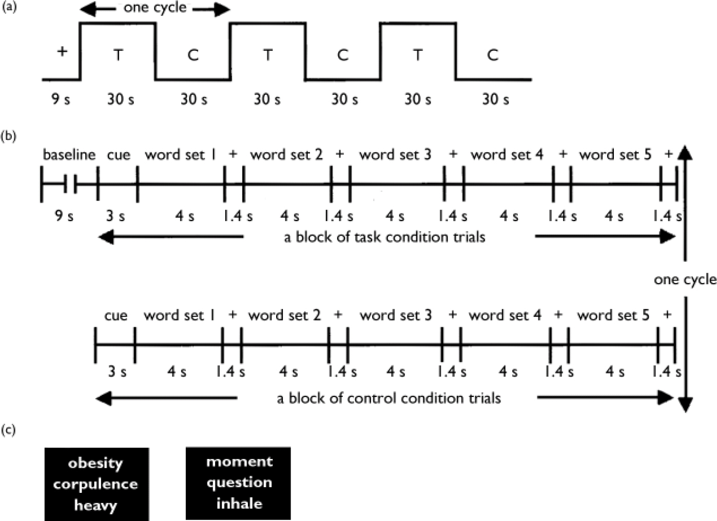
Fig. 1 Design of the study task. (a) Six alternating blocks of task condition (T) trials and control condition (C) trials were presented successively; the total scan time was 189 s (3 min and 9 s), yielding 63 images of 28 axial slices (1764 images). (b) Blocks of task condition and control condition trials were preceded by a baseline imaging period. Each block began with a cue (‘task’ or ‘control’). The participant selected the word judged to be the most unpleasant or most neutral in each word set, by pressing one of three buttons. (C) Translations of typical word sets presented in this study (left-hand block, task condition; right-hand block, control condition).
Participants were instructed to select the most unpleasant word from each set of unpleasant words based on their personal knowledge and experience, and for each set of neutral words, participants were instructed to select the word that they thought was the most neutral; they indicated their choice by pressing one of three buttons on a response pad in the MRI scanner.
Image acquisition and processing
The MRI scanner used was a Magnex Eclipse 1.5 T Power Drive 250 (Shimadzu Medical Systems, Kyoto, Japan). A time-course series of 63 volumes was acquired with T 2 *-weighted, gradient echo, echo planar imaging (EPI) sequences. Each volume consisted of 28 slices, each 4.0 mm thick with no gap, encompassing the entire brain. The interval between two successive acquisitions of the same image (time to repetition, TR) was 3000 ms, the time to echo (TE) was 55 ms and the flip angle was 90°. The field of view was 256 mm and the matrix size 64 × 64, giving voxel dimensions of 4.0 mm × 4.0 mm × 4.0 mm. After fMRI scanning, structural scans were acquired using a T 1-weighted gradient echo pulse sequence (TR 12 ms, TE 4.5 ms, flip angle 20°, field of view 256 mm, voxel dimensions 1.0 mm × 1.0 mm × 1.0 mm), to facilitate localisation and co-registration of the functional data.
Image processing and statistical analysis were performed using Statistical Parametric Mapping (SPM) 99 software (Wellcome Department of Cognitive Neurology, London, UK) implemented in Matlab (Mathworks, Inc., Natick, MA, USA). The first two volumes of the fMRI run (pre-task period) were discarded because the magnetisation was unsteady, and the remaining 61 volumes were used for the statistical analysis. Images were corrected for motion and realigned with the first scan of the session, which served as the reference. The T 1 anatomical images were co-registered to the first functional images in each individual and aligned to a standard stereotaxic space, using the Montreal Neurological Institute (MNI) T 1 template in SPM99. The calculated non-linear transformation was applied to all functional images for spatial normalisation. Finally, the fMRI images were smoothed with a 12 mm full-width, half-maximum Gaussian filter.
Using group analysis according to a random effect model that allowed inference to the general population (Reference Friston, Holmes and WorsleyFriston et al, 1999), we first identified brain regions that showed a significantly greater response to unpleasant word sets in comparison with the response to neutral word sets among male and among female participants, as brain areas related to the cognition of unpleasant word stimuli concerning body image in men and women, respectively. We then took the data of 13 of the 15 women who had participated in our previous study (Reference Shirao, Okamoto and OkadaShirao et al, 2003a ) and directly compared the activation of the entire brain in the male and female sub-samples using the two-sample Student's t-test. The resulting set of voxel values for each contrast constituted an SPM{t} map. The SPM{t} maps were then interpreted by referring to the probabilistic behaviour of Gaussian random fields. The data were given an initial threshold at an uncorrected P < 0.001 at the voxel level, and regions about which we had an a priori hypothesis were reported at this threshold (Reference Elliott, Friston and DolanElliott et al, 2000). For regions about which there was no clear hypothesis, a more stringent threshold of P < 0.05 corrected at the cluster level of multiple comparison was used. The x, y and z coordinates provided by SPM, which were in MNI brain space, were converted to the x, y and z coordinates in Talairach & Tournoux's (TT) brain space (Reference Talairach and TournouxTalairach & Tournoux, 1988) using the following formulae:
Labels for brain activation foci were obtained in Talairach coordinates using the Talairach Daemon software (Research Imaging Center, University of Texas, TX, USA), which provides accuracy similar to that of neuroanatomical experts (Reference Lancaster, Woldorff and ParsonsLancaster et al, 2000). The labelling of areas given by this software was then confirmed by comparison with activation maps overlaid on MNI-normalised structural images.
Evaluation of pleasantness and familiarity of the word stimuli
Each participant was asked to rate the pleasantness and familiarity of all the words presented in the tasks on a scale from 1 (very unfamiliar; very unpleasant) to 7 (very familiar; very pleasant), immediately after scanning. For this rating procedure the list of words was presented in randomised order in a table format.
RESULTS
Rating of words
The ratings of familiarity with the two categories of words did not significantly differ among men (mean familiarity score: unpleasant words 3.8, neutral words 4.4, P=0.054 by two-tailed Wilcoxon single-rank test) or women (mean familiarity score: unpleasant words 4.3, neutral words 4.3, P=0.456). However, all participants rated the unpleasant words concerning body image as significantly more unpleasant than the neutral words (mean pleasantness score: unpleasant words 3.1, neutral words 4.1, P=0.007 in men; unpleasant words 2.7, neutral words 4.1, P=0.002 in women). Neither the ratings of pleasantness nor the ratings of familiarity in each word category significantly differed between the male and female groups.
Brain activation
In men there was significantly greater activation of the left hippocampus, left superior temporal gyrus, left fusiform gyrus and left medial frontal gyrus when the emotional decision task involved unpleasant words compared with neutral words, whereas the women showed significantly greater activity of the left parahippocampal gyrus including amygdala, left thalamus and right caudate body in the same comparison (Table 1, Fig. 2).
Table 1 Relative increases in brain activity associated with unpleasant words concerning body image (task) and neutral words (control)
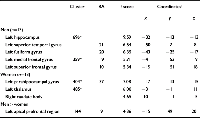
| Cluster | BA | t score | Coordinates1 | |||
|---|---|---|---|---|---|---|
| x | y | z | ||||
| Men (n=13) | ||||||
| Left hippocampus | 696* | 9.59 | -32 | -13 | -13 | |
| Left superior temporal gyrus | 21 | 6.54 | -50 | -7 | -8 | |
| Left fusiform gyrus | 20 | 6.35 | -43 | -25 | -17 | |
| Left medial frontal gyrus | 359* | 9 | 5.71 | -4 | 53 | 9 |
| Left superior frontal gyrus | 10 | 5.34 | -15 | 51 | 18 | |
| Women (n=13) | ||||||
| Left parahippocampal gyrus | 404* | 37 | 7.08 | -17 | -13 | -15 |
| Left thalamus | 485* | 6.08 | -3 | -11 | 11 | |
| Right caudate body | 4.65 | 10 | 1 | 5 | ||
| Men > women | ||||||
| Left apical prefrontal region | 144 | 9 | 4.36 | -15 | 49 | 20 |

Fig. 2 Brain areas showing significantly greater activation during the task condition compared with the control condition. Three-dimensional ‘look-through’ projections of statistical parametric maps of the brain regions are shown (one-sample Student's t-test; corrected P < 0.05 at the cluster level; n=13; d.f.=12).
The two-sample Student's t-test revealed that there was a significantly higher BOLD response in the left apical prefrontal region in men than in women during the unpleasant word task compared with neutral word task (Table 1, Fig. 3). No brain area showed significantly higher activation in women than in men during any of the tasks.

Fig. 3 Brain regions showing significantly greater activation in men than in women during the task condition of the emotional decision task compared with the control condition. Clusters of activation are overlaid onto a T 1-weighted anatomical magnetic resonance image. The white spots show areas of high activation. Two-sample Student's t-test; uncorrected P < 0.001 in height; n=26 (13 men, 13 women); d.f.=24.
Correlation between psychological data and brain activation
Among the 13 women participants, activation in the left apical prefrontal area, which was significantly lower than that in men during the unpleasant words task, was negatively correlated with the total EDI–2 score (Spearman's rank-order correlation analysis: correlational coefficient -0.699, P=0.008). There was no correlation between any brain area showing significant BOLD response and the EDI–2 scores or the pleasantness rating of the unpleasant words.
DISCUSSION
We used the emotional decision task to examine the brain areas engaged in the perception of unpleasant words concerning body image and to compare the patterns of brain activation in men and women. Our results showed that the left medial part of the frontal gyrus, the left limbic area excluding the amygdala, the left superior temporal gyrus and the left fusiform gyrus play an important part in processing unpleasant words concerning body image in men.
Lack of amygdalar activation in men
Consistent with our hypothesis, the amygdala did not show significant activation among men; however, the gender difference of the BOLD response in the amygdala was not significant by two-sample Student's t-test.
The amygdala has been suggested by many studies to be strongly associated with stimuli signalling threat. Human lesion and imaging studies consistently indicate that the amygdala is concerned in fear conditioning (Reference Morris, Ohman and DolanMorris et al, 1998), in the recognition of fearful facial expressions (Reference AdolphsAdolphs, 1999) and in the evocation of fearful emotional responses from direct stimulation (Reference Halgren, Walter and CherlowHalgren et al, 1978). The amygdala is also considered to be important in the detection of environmental threat (Reference Scott, Young and CalderScott et al, 1997), including verbal stimuli (Reference Isenberg, Silbersweig and EngelienIsenberg et al, 1999). Therefore, the lack of significant activation in the amygdala among men suggests that men may not process unpleasant words concerning body image as fearful information, whereas women seem to do so.
Medial prefrontal cortex and emotional processing
The significant activation in the medial part of the frontal gyrus – Brodmann areas (BAs) 9 and 10; medial prefrontal cortex – was only detected in men, and there was a significantly higher BOLD response in men than in women in the left apical prefrontal region (BA 9) when performing the unpleasant word task compared with the neutral word task by two-sample Student's t-test. These results were consistent with our hypothesis. Many previous studies have suggested that the medial prefrontal cortex might have a role generally in emotional processing. It is reported that visual stimuli that evoke emotions, such as films or pictures, activated the medial prefrontal cortex, and that recall of various emotions such as happiness, sadness and disgust, and a mixture of these emotions, all separately engaged this brain region (Reference Lane, Reiman and BradleyLane et al, 1997; Reference Reiman, Lane and AhernReiman et al, 1997). Several more recent studies suggest that when people turn their attention inwards to assess self-relevant attributes or emotional awareness, activity increases in the medial prefrontal cortex (Reference Johnson, Baxter and WilderJohnson et al, 2002; Reference Zysset, Huber and FerstlZysset et al, 2002). The medial prefrontal cortex has connections to limbic structures, including the amygdala, constituting an interaction zone between emotional processing and cognitive processing (Reference Drevets and RaichleDrevets & Raichle, 1998), and this region may have a role in modulating the emotional response in the amygdala and other limbic structures. Limbic structures, including the amygdala, are likely to respond to emotional stimuli at a sensory or perceptual level (Reference Reiman, Lane and AhernReiman et al, 1997), whereas the medial prefrontal cortex may be involved in the cognitive aspects of emotional processing, such as attention to emotion, appraisal or identification of emotion (Reference Drevets and RaichleDrevets & Raichle, 1998). From this viewpoint, the gender differences detected in our study may demonstrate differences of cognitive pattern in men and women. Our results suggest the possibility that men processed the emotional decision task including words concerning body image more cognitively rather than emotionally, and activation in the medial prefrontal cortex was prominent; on the other hand, women processed this task more emotionally rather than cognitively, and the medial prefrontal cortex did not exhibit any significant activation. Both men and women perceived the unpleasantness of the words concerning body image to the same degree, according to their subjective ratings, but the fMRI data suggest that their processes are different: women are likely to use more intuitive processing whereas men use more rational processing. This discrepancy between the genders in cognitive style related to body image may contribute to the large gender difference in susceptibility to eating disorders.
Another possible explanation of the different patterns of activation in the medial prefrontal cortex between men and women may be the difference in men's familiarity with the unpleasant word set compared with the neutral words. Although the ratings of familiarity were not different between men and women (P=0.133 by Mann–Whitney U test), there was a trend for male participants to be less familiar with the unpleasant words concerning body image than with the neutral words (P=0.054 by two-tailed Wilcoxon single-rank test). When processing unfamiliar words concerning body image, men might turn more attention inwards, and subsequently the BOLD response in the medial prefrontal cortex was higher than while processing neutral words.
Among women, correlational analysis revealed that the BOLD response in the left apical prefrontal region (BA 9), which was significantly lower in women than in men, was negatively correlated with total EDI–2 scores; in other words women with higher EDI–2 scores exhibited lower activity in this brain area. These results suggest the possibility that the apical prefrontal region might be involved in the pathophysiology of eating disorders.
Comparison with other neuroimaging studies
To our knowledge, two fMRI studies concerning body image distortion have investigated the effects of pictorial body image stimuli in women with anorexia nervosa and healthy controls (Reference Seeger, Braus and RufSeeger et al, 2002; Reference Wagner, Ruf and BrausWagner et al, 2003). One study reported that patients with anorexia nervosa showed activation in the right amygdala, right fusiform gyrus and brain-stem associated with stimulation with their own body image whereas healthy controls showed activation only in the fusiform gyrus (Reference Seeger, Braus and RufSeeger et al, 2002), and the other reported that patients with anorexia nervosa showed greater activation in the prefrontal cortex and the inferior parietal lobule than did controls (Reference Wagner, Ruf and BrausWagner et al, 2003). The latter authors explain the discrepancy between these results as a consequence of the design of the task. Many differences in the experimental conditions between these studies and ours make it difficult to compare the brain activation data, but a possible explanation of the discrepancy between the study by Wagner et al (Reference Wagner, Ruf and Braus2003) and our study is the age of the participants: those in the former study were adolescents (approximately 15 years old), whereas we recruited young adults (approximately 25 years old). An fMRI study which investigated the brain activation of adult and adolescent men and women while processing emotional facial expressions reported that the adult men and adolescents (both boys and girls) showed significant activation in the bilateral orbitofrontal cortex and anterior cingulate cortex in response to an angry face, whereas the adult women showed significant activation in the left amygdala in addition to these brain areas (Reference McClure, Monk and NelsonMcClure et al, 2004). These results suggest that the patterns of neural responses to emotional stimuli may be different in adults and adolescents.
A positron emission tomography study of gender differences in brain activation patterns during recognition of emotional facial expressions revealed that greater amygdalar activation was observed in women and greater medial frontal activation was observed in men (Reference Hall, Witelson and SzechtmanHall et al, 2004); these authors suggest that men might take a more analytic approach and might regulate their emotional reaction to the stimuli more than women. Although the categories of stimuli are different, these results support our findings.
Study limitations
Our study has some limitations. First, we did not administer a structured interview when selecting the participants; however, they had no psychiatric or neurological illness at the time of their participation, although we cannot rule out its occurrence in the future. Second, participants were asked to rate only the unpleasantness and familiarity of the words used. If we had also asked about the fearfulness induced by the stimuli, we might have found gender differences in subjective rating and the results with brain image data would have been more clear-cut. Last, although our data suggest that there is differential activation of the brains of men and women when processing unpleasant words concerning body image, we cannot conclude whether these results are specific to unpleasant stimuli concerning body image or would apply to a wide range of unpleasant stimuli. Among women, a lower BOLD response in the prefrontal region compared with men while processing unpleasant words concerning body image exhibited a negative correlation with the total EDI–2 score, but it is unclear whether this brain region is the focal area responsible for susceptibility to eating disorders.
In conclusion, our study revealed that the paralimbic area including the amygdala was activated only in women and that the left medial prefrontal cortex was activated only in men while performing the emotional decision task with unpleasant words concerning body image. These results suggest that gender differences in brain activation might explain the differences in the style of cognition towards unpleasant stimuli concerning body image. Further studies comparing people who have eating disorders with healthy controls and which include general unpleasant word stimuli to contrast with words specific to body image are needed to elucidate the neural substrate responsible for the onset of eating disorders.
Clinical Implications and Limitations
CLINICAL IMPLICATIONS
-
▪ Gender differences in brain activation suggest differences between men and women in the style of cognition toward unpleasant stimuli concerning body image.
-
▪ This discrepancy in cognitive style may have relevance to the large gender difference in susceptibility to eating disorders.
-
▪ The medial prefrontal cortex may be the brain area linked to the pathophysiology of eating disorder.
LIMITATIONS
-
▪ We did not use a structured interview when selecting participants.
-
▪ We asked the participants to rate only pleasantness and familiarity of the word stimuli and we could find no clear relationship between brain activation and the subjective rating of the words concerning body image.
-
▪ It is unclear whether the patterns of activation in the prefrontal area were specific to the stimuli concerning body image.
Acknowledgement
The study was supported by the Research on Psychiatric and Neurological. Diseases and Mental Health, Ministry of Health, Labour and Welfare, Japan.


eLetters
No eLetters have been published for this article.